
In-office CBCT • Downtown Honolulu
See your teeth and jaw in 3D
How close is a wisdom tooth to a nerve? Is there enough bone for an implant? When a dental X-ray leaves a question open, our KaVo OP 3D scanner lets us look at the area from another angle, right here on Bethel Street.
Request an appointment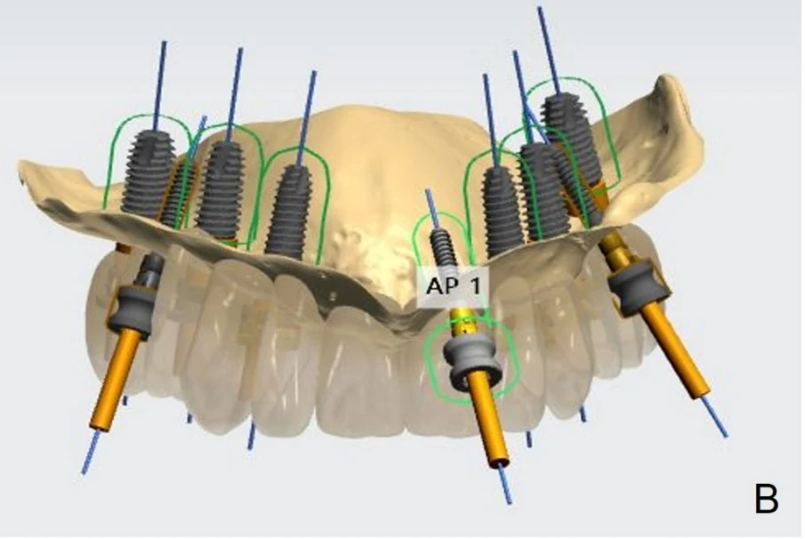
Inside a 3D dental scan
What can we see beneath the gums?
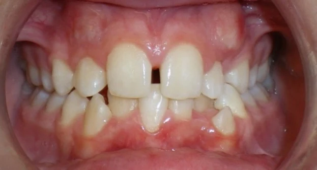
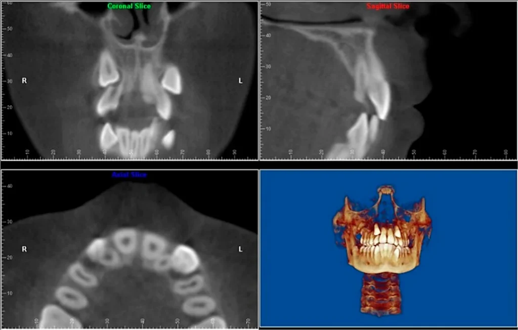
Cone beam computed tomography (CBCT) uses a rotating X-ray beam to build a three-dimensional picture of your teeth and bone. We can turn that picture and look through it one thin slice at a time. The photo and scan below show the same person.
Wisdom tooth removal
How close are the roots to the nerve?
A lower wisdom tooth can overlap the nerve canal on a panoramic X-ray. If the answer could change treatment, a CBCT scan can show whether the canal passes alongside the roots or beneath them. This nerve carries feeling to your lower lip and chin.
That view helps us explain the risks of removal and decide whether to involve an oral surgeon. When roots lie very close to the canal, the surgeon may discuss removing just the crown and leaving the roots in place.
Explore oral surgery at our office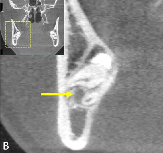
Dental implant planning
Plan the implant around the tooth it will support
A replacement tooth needs to meet your bite and have bone beneath it for support. We can measure the width and height of your jawbone, map the sinus or nerve canal nearby, and compare implant positions before surgery. If bone is thin, we can discuss a graft while we plan your care.
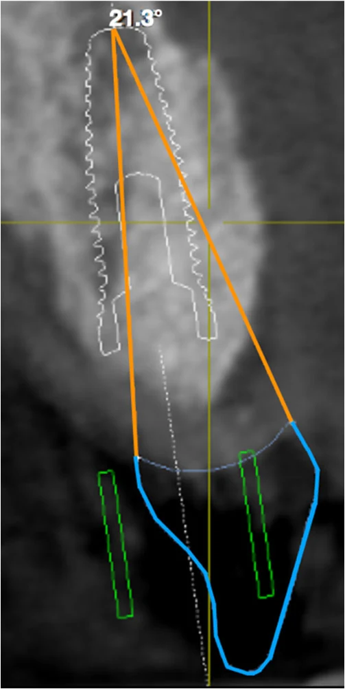
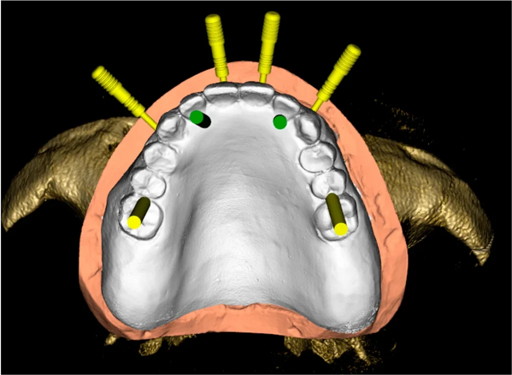
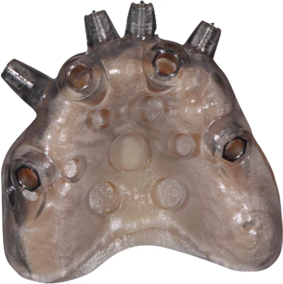
When the source of tooth pain is unclear
Sometimes the examination and standard X-rays leave more than one possible explanation for symptoms. A focused scan can help us decide where to look next.
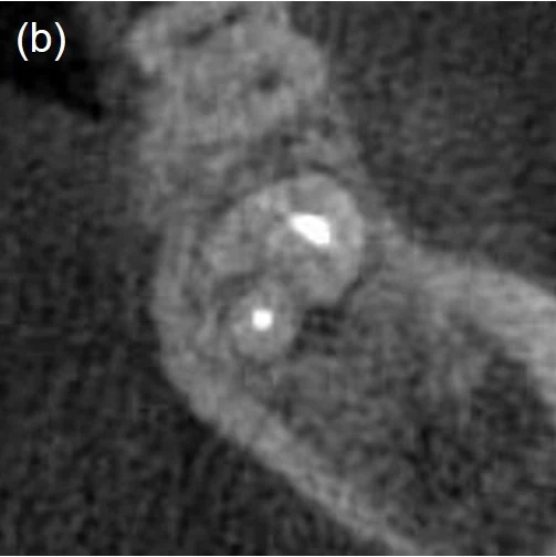
Your visit on Bethel Street
What it’s like to have a scan here
- Before the scan, we explain the question we’re trying to answer. We’ll help position your head and may ask you to remove glasses or jewelry.
- During the scan, you hold still while the open arm moves around your head. The scan itself takes seconds and is painless.
- After the scan, we can bring the images up in our office, point out what we see, and talk through the next step. Having the scanner here can save you a separate trip to an imaging center.
Questions patients ask
Will I need a 3D scan at my checkup?
Most routine dental questions can be answered with an exam and standard X-rays. We recommend CBCT when the extra detail is likely to change a decision about your care.
How much radiation does a CBCT scan use?
A dental CBCT scan usually uses more radiation than a standard dental X-ray. The dose depends on the area and settings. We select the smallest area that answers your question and tailor the settings to you. Please tell us if you are pregnant or might be pregnant.
What if I already have a scan from another office?
Let us know when you schedule. We can help you request the original scan and review whether it covers the area we need before recommending another one.
Have a question about your X-ray?
Bring it to your visit. We’ll look at it with you and explain whether a 3D scan would change your options.
Request an appointment
Clinical image credits and patient resources
The clinical pictures on this page are published teaching examples. The consultation room photograph is from A Downtown Dental Group. The clinical images were resized or converted to WebP, and individual panels were extracted where appropriate. Reproduced under Creative Commons Attribution 4.0.
- Upper-jaw implant plan — Seung Wook Jung, Yi-Qin Fan and Chunui Lee, 2022. Figure 3B.
- Clinical photograph and scan of unerupted canines — Ahmad Abdelkarim, 2019. Figures 1 and 3.
- Wisdom tooth and nerve canal — Adib Al-Haj Husain and colleagues, 2023. Figure 3B.
- Single implant and planned crown — Mario Caggiano and colleagues, 2022. Figure 1a.
- Full-arch implant plan and surgical guide — Francesca Cattoni and colleagues, 2021. Figures 5 and 7b.
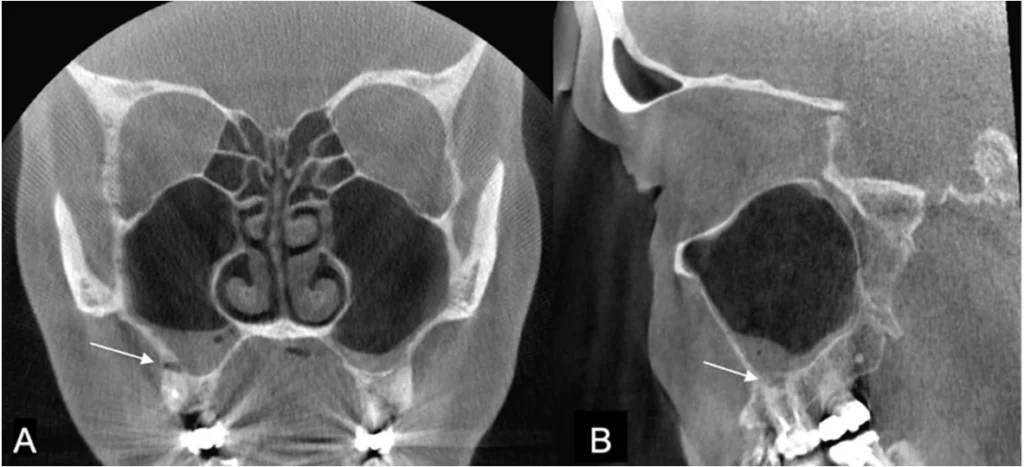
- Molar roots and sinus — Niina Kuusisto and colleagues, 2023. Figure 2.
- Previously treated root canal — Da-Min Park, Woo-Hyun Seok and Ji-Young Yoon, 2024. Figure 3b.
Dental cone beam CT for patients · FDA information on dental CBCT · ADA and AAOMR imaging recommendations